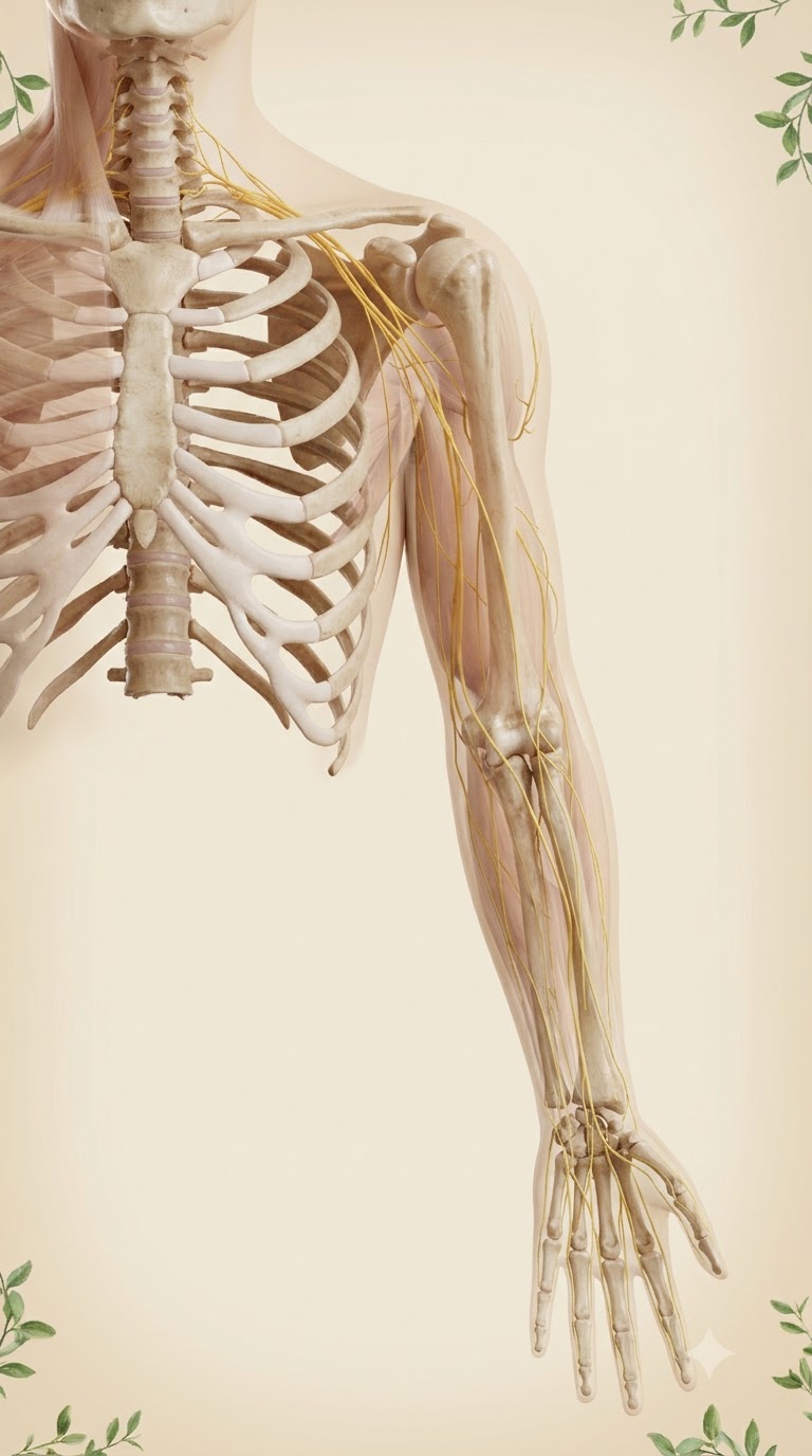
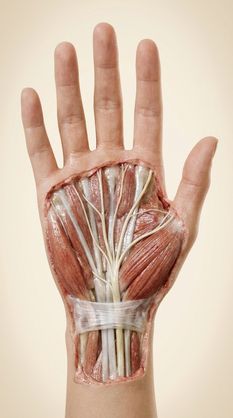
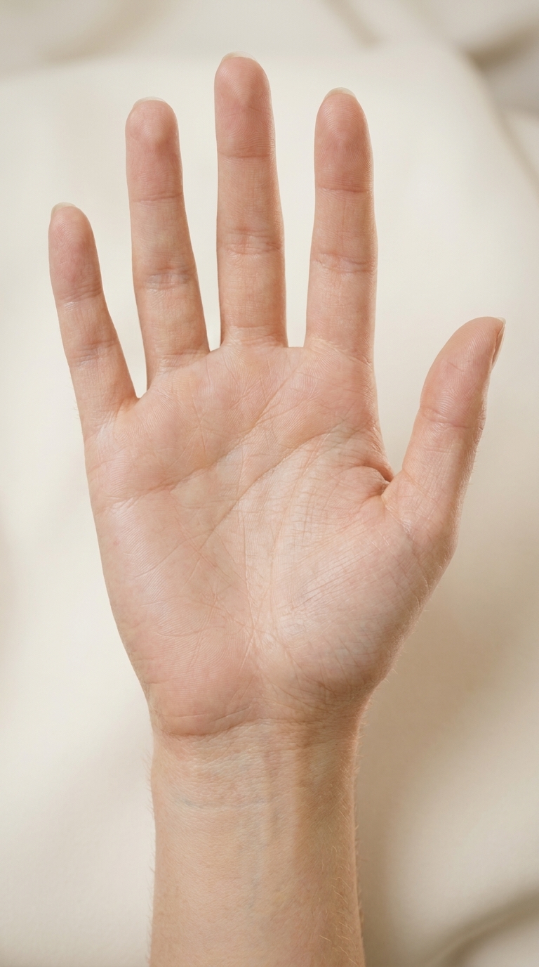


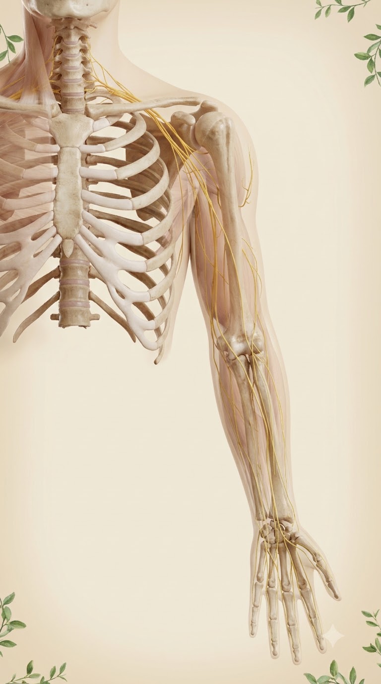






Use the reference image (VOKA 3D anatomy atlas: nerves of the right upper limb) as the exact anatomical guide. Photorealistic 3D medical anatomy render, premium 3D atlas quality, anatomically correct skeleton. Front (anterior) view of the right half of the neck and upper chest and the whole right arm down to the fingertips; arm hanging along the body, palm facing forward, thumb pointing away from the body. Skeleton in ivory: cervical spine in the neck, sternum and upper ribs of the chest; the clavicle runs horizontally from the sternum (manubrium) at the midline out to the acromion of the shoulder — it does NOT attach to the spine; scapula behind the ribs; humerus, radius, ulna, carpal bones and five fingers. Nerves in warm yellow, realistic texture: roots C5–T1 emerge from the side of the cervical spine, form the brachial plexus that passes behind the clavicle into the armpit, then the median nerve runs down the inner side of the arm, crosses the front of the elbow, goes down the middle of the forearm, through the wrist into the palm to the thumb, index, middle and half of the ring finger; the ulnar nerve goes behind the inner elbow to the little finger; the radial nerve winds behind the humerus to the back of the forearm. Muscles shown faint and semi-transparent so the nerves are clearly visible. Soft warm studio light, clean, physiological, not gory. Background: plain warm cream tone with soft vignette, a couple of small green leafy sprigs in the corners. No text, no labels, no watermark. Vertical 9:16, everything from the neck to the fingertips fits in the frame.
Бесплатный старт · Генерация видео и картинок с ИИ за секунды